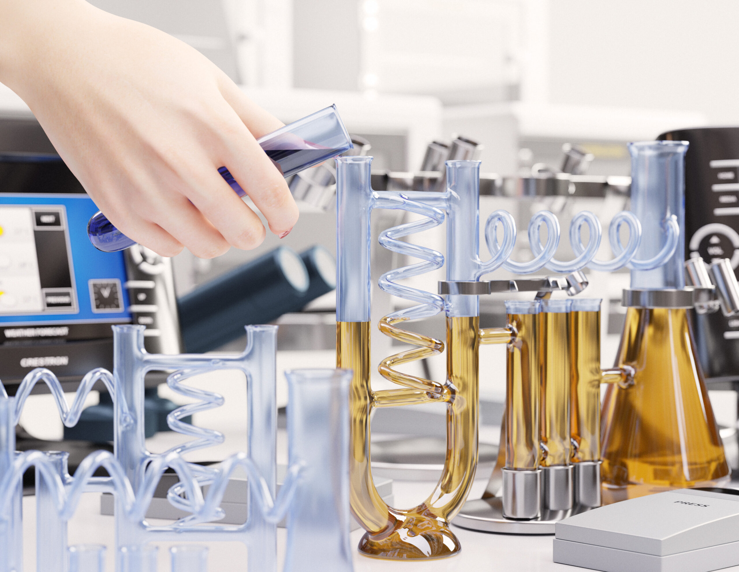
An aptamer library is a diverse pool of nucleic acid sequences (DNA or RNA) from which specific aptamers—short oligonucleotides that bind to target molecules with high affinity—can be selected. Constructing a high-quality library is the foundation of aptamer screening technologies like SELEX. 2. Key Components of an Aptamer Library Randomized Region The central portion of the aptamer, typically 20–60 nucleotides, is randomized to generate diversity. Example: N20–N40 where N = A, T/U, G, or C. The diversity determines the probability of finding high-affinity binders. Flanking Constant Regions Short sequences (~15–25 nt) at both ends of the randomized region. Functions: Primer binding sites for PCR amplification. Stability and structural constraints. Overall Length Usually 40–100 nucleotides, balancing structural complexity and amplification efficiency. 3. Steps of Library Construction Design of Oligonucleotides Include random regions flanked by known primer sequences. Example structure:5'-[Primer]-N40-[Primer]-3' Chemical Synthesis Use solid-phase DNA/RNA synthesis to generate the oligonucleotides. Random nucleotides are incorporated using a controlled mixture of A, T/U, G, C. Amplification (for DNA libraries) PCR amplifies the synthesized sequences. RNA libraries require in vitro transcription from DNA templates. Purification Remove truncated or incomplete sequences. Methods: PAGE purification or HPLC. Quality Control Ensure correct length, diversity, and absence of biases.…

Core Technology: SELEX The foundation of all these services is the SELEX process, an in vitro method to select aptamers from a vast random library (typically 10^13 - 10^15 unique sequences). The library is incubated with the target, unbound sequences are washed away, and bound sequences are eluted and amplified by PCR (for DNA) or RT-PCR (for RNA). This cycle is repeated 8-15 times to enrich for the tightest binders. Services for Protein Targets This is the most common application, as aptamers are often touted as "chemical antibodies." 1. Standard Protein SELEX: Target: Purified, recombinant proteins (e.g., cytokines, receptors, enzymes, viral capsids). Key Considerations: Protein Purity & Conformation: Critical for success. Services often require >90% purity and verification of native folding. Immobilization: The protein is usually immobilized on beads (e.g., streptavidin/biotin, Ni-NTA/His-tag) to facilitate partitioning. Some services offer solution-phase SELEX to avoid conformation changes. Counter-Selection: To ensure specificity, libraries are pre-incubated with related proteins or the immobilization matrix to subtract non-specific binders. 2. Specialized SELEX for Complex Proteins: Membrane Protein SELEX: For receptors and channels. Requires special handling (e.g., use of nanodiscs, detergent micelles, or whole cells overexpressing the target). Post-Translationally Modified Protein SELEX: For targets where phosphorylation, glycosylation, etc., are essential for function. 3. Cell-SELEX (for Cell-Surface…
What is Protein SELEX? SELEX (Systematic Evolution of Ligands by EXponential Enrichment) is an iterative, in vitro process used to discover aptamers—single-stranded DNA or RNA molecules that bind to a specific target (like a protein) with high affinity and specificity. Protein SELEX specifically refers to using a purified protein as the target to isolate aptamers against it. These aptamers are often called "chemical antibodies" due to their similar binding function. Core Workflow of a Protein SELEX Service A professional service will manage this entire complex process, typically involving the following stages: 1. Project Design & Consultation Target Characterization: Discussing the target protein's properties (size, purity, stability, domains, post-translational modifications). Selection Strategy: Choosing the right SELEX variant (e.g., Nitrocellulose filter, Magnetic bead, Capillary Electrophoresis, or Cell-SELEX for membrane proteins). Defining counter-selection steps to avoid binders to unwanted tags or impurities. Library Design: Using a standard or custom random oligonucleotide library (e.g., 40-60 random nucleotides flanked by primer sites). 2. The SELEX Cycle (Repeated 8-15 Rounds) mermaid graph TD A[Start: ssDNA/RNA Library<br>~10^15 unique sequences] --> B{Incubation with<br>Target Protein}; B --> C[Partition: Separate<br>Bound from Unbound Sequences]; C --> D[Elution: Recover<br>Bound Sequences]; D --> E[Amplification:<br>PCR (DNA) or RT-PCR (RNA)]; E --> F[Purification:<br>Regenerate ssDNA/RNA for next round]; F --> G{Enrichment<br>Sufficient?}; G -- No…
Antibody: A large, Y-shaped protein produced naturally by the immune system (B cells) in response to a foreign substance (antigen). It is a biological molecule. Aptamer: A short, single-stranded piece of DNA or RNA (or modified nucleotides) that is artificially engineered in a lab to bind to a specific target. It is a chemical molecule. Key Differences at a Glance Feature Antibody Aptamer Chemical Nature Protein (IgG, etc.) Nucleic Acid (DNA or RNA) Origin Biological (from animals) Chemical (SELEX process in vitro) Size Large (~150 kDa) Small (~10-30 kDa) Production Requires animal immunization or cell culture. Batch-to-batch variability possible. Synthetic, produced by chemical synthesis. Highly reproducible. Targets Primarily immunogenic targets (proteins, pathogens). Limited to targets that elicit an immune response. Extremely broad: ions, small molecules, proteins, cells, viruses, tissues. Can target toxins or non-immunogenic substances. Stability Sensitive to temperature (often requires refrigeration), pH, and proteases. Can denature. Thermally stable, can be renatured after denaturation. Resistant to harsh conditions (pH, organic solvents). Modification Difficult to modify chemically without affecting function. Site-specific conjugation is complex. Easy to chemically modify with reporters, drugs, or linkers at precise locations. Immunogenicity Can itself trigger an immune response (especially non-human antibodies). Generally low immunogenicity, but can be designed to be non-immunogenic. Cost…

1. Therapeutics & Medicine This is one of the most promising areas. Drugs: The first FDA-approved aptamer drug is Pegaptanib (Macugen) for treating age-related macular degeneration. It binds to VEGF, a protein that promotes abnormal blood vessel growth. Targeted Drug Delivery: Aptamers can be attached to drug nanoparticles or toxins, acting as a "homing device" to deliver the payload specifically to cancer cells or diseased tissues, minimizing side effects. Antidotes: "Antidote" or control oligonucleotides can be designed to bind and deactivate an aptamer's function, allowing for precise control of therapeutic activity—something very difficult with antibodies. Antiviral & Antibacterial Agents: They can bind to and neutralize viruses (like HIV, influenza, SARS-CoV-2) or specific bacterial proteins. 2. Diagnostics & Biosensing Aptamers are powerful tools for detecting molecules. Aptamer-based Assays: Used in ELISA-like formats (sometimes called ELASA) to detect biomarkers for diseases (cancer, infections) in blood or other samples. Point-of-Care Tests: Integrated into portable biosensors (aptasensors) for rapid, on-site detection of pathogens, toxins, or hormones. They can use optical, electrochemical, or mass-sensitive methods. Medical Imaging: Labeled with fluorescent dyes or radioisotopes, aptamers can help visualize tumors or diseased tissues during surgery or in scans. 3. Research & Biotechnology Protein Function Studies: Used to inhibit specific proteins in cells or in vitro to study their biological function, similar to using…
Aptamers are single-stranded DNA or RNA oligonucleotides (typically 20-80 bases) that fold into specific 3D structures capable of binding target molecules with high affinity and specificity, earning them the nickname "chemical antibodies." Their unique properties make them promising agents for targeted liver cancer therapy. Why Aptamers Are Suitable for Liver Cancer Targeting Molecular Recognition Capabilities Can be selected against specific liver cancer biomarkers (ASGPR, GPC3, EGFR, etc.) High binding affinity (nM to pM range) Specific discrimination between cancerous and normal hepatocytes Advantages Over Antibodies Smaller size (5-25 kDa) for better tissue penetration Chemical synthesis without batch variation Lower immunogenicity Easier modification and conjugation Higher thermal stability Key Targeting Strategies for Liver Cancer 1. Targeted Drug Delivery Aptamer-drug conjugates: Direct conjugation of chemotherapeutic agents (doxorubicin, sorafenib derivatives) Nano-carrier guidance: Aptamers decorating nanoparticles, liposomes, or micelles containing drugs Targeted prodrug activation: Aptamer-mediated delivery of enzyme prodrug systems 2. Targeted Gene Therapy Delivery of siRNA/miRNA to regulate oncogene expression CRISPR/Cas9 delivery for gene editing Examples: Anti-GPC3 aptamers delivering VEGF siRNA to inhibit angiogenesis 3. Multifunctional Theranostic Applications Combined imaging (fluorescence, PET, MRI) and therapy Aptamer-conjugated agents for image-guided surgery or ablation 4. Immomodulation Targeting immune checkpoint molecules (PD-1/PD-L1) Redirecting immune cells to tumor sites Clinically Relevant Targets…

Why Aptamers are Promising for Liver Cancer Imaging Compared to traditional antibodies, aptamers offer key advantages for in vivo applications: Small Size (5-15 kDa): Enables better tissue penetration and faster blood clearance, leading to higher tumor-to-background ratios. Low Immunogenicity: Reduced risk of allergic reactions or neutralization upon repeated administration. Ease of Chemical Synthesis & Modification: Can be stably produced, and easily conjugated with dyes, radionuclides, or nanoparticles. Rapid Tissue Penetration & Clearance: Ideal for imaging shortly after injection. Engineerable Flexibility: Can be designed as multivalent or bispecific constructs. Key Steps in Developing Aptamers for Liver Cancer Imaging Target Selection: Identifying a molecule highly expressed on liver cancer cells but low on normal hepatocytes is critical. Prime targets include: Glypican-3 (GPC3): A heparan sulfate proteoglycan overexpressed in 70-80% of hepatocellular carcinomas (HCC). Alpha-fetoprotein (AFP): A classic serum biomarker, with membrane-bound forms also present on HCC cells. Epithelial Cell Adhesion Molecule (EpCAM): Expressed on cancer stem cells in HCC and cholangiocarcinoma. Asialoglycoprotein Receptor (ASGPR): Highly expressed on normal hepatocytes but often dysregulated in HCC; useful for "background" subtraction or targeting specific isoforms. Receptor Tyrosine Kinases: Like c-Met or VEGFR2. Aptamer Generation: Typically done via SELEX (Systematic Evolution of Ligands by EXponential enrichment). For liver cancer, Cell-SELEX using live HCC cells vs. normal hepatocytes is preferred, as it identifies aptamers…

Aptamers are emerging as powerful molecular tools for the in vitro detection of liver cancer, offering a promising alternative to traditional antibodies. Here’s a comprehensive breakdown: What are Aptamers? Aptamers are short, single-stranded DNA or RNA oligonucleotides (or peptides) that bind to specific target molecules (proteins, cells, small molecules) with high affinity and specificity. They are selected in vitro through a process called SELEX (Systematic Evolution of Ligands by Exponential Enrichment). Why Aptamers for Liver Cancer Diagnosis? Compared to conventional antibodies, aptamers offer key advantages for diagnostics: High Specificity & Affinity: Can distinguish between healthy and cancerous biomarkers. Small Size: Better tissue penetration and access to epitopes. In Vitro Synthesis: Chemically produced, resulting in low batch-to-batch variation. Stability: Thermally stable and easily modifiable. Non-Immunogenic: Suitable for repeated use in assays. Key Targets for Liver Cancer Detection Aptamers are developed to detect liver cancer (primarily Hepatocellular Carcinoma, HCC) by targeting: Circulating Protein Biomarkers: Alpha-fetoprotein (AFP): The most widely used serum biomarker for HCC, but with limited sensitivity/specificity. AFP-specific aptamers are used in electrochemical, fluorescent, and colorimetric sensors to improve detection limits. Glypican-3 (GPC3): A cell-surface proteoglycan overexpressed in >70% of HCCs. GPC3 aptamers are central to many sensitive detection platforms. Vascular Endothelial Growth Factor (VEGF): Associated with angiogenesis and metastasis. Platelet-Derived Growth Factor (PDGF): Involved in…

The application of aptamers in the targeted diagnosis of liver cancer, particularly Hepatocellular Carcinoma (HCC), represents a cutting-edge frontier in oncology and molecular diagnostics. Aptamers offer a powerful alternative to traditional antibodies, with the potential to revolutionize early detection, imaging, and personalized treatment. Here is a comprehensive breakdown of their applications: 1. What are Aptamers? Aptamers are single-stranded DNA or RNA oligonucleotides (or peptides) that bind to specific target molecules (proteins, cells, small molecules) with high affinity and specificity. They are often termed "chemical antibodies." They are selected in vitro through a process called SELEX (Systematic Evolution of Ligands by EXponential enrichment). Key Advantages Over Antibodies: Small size: Better tissue penetration. Chemical synthesis: Highly reproducible, no batch-to-batch variation. Stability: Thermally stable and can be reversibly denatured. Low immunogenicity: Unlikely to provoke an immune response. Ease of modification: Can be easily labeled with dyes, radioisotopes, or nanoparticles. 2. Core Applications in Liver Cancer Diagnosis A. Detection of Circulating Biomarkers (Liquid Biopsy) This is the most prominent application. Aptamers are used as capture/detection probes in biosensors to identify HCC-specific biomarkers in blood, enabling non-invasive, early diagnosis. Targeting Protein Biomarkers: Alpha-fetoprotein (AFP): The most common clinical serum marker for HCC, but its sensitivity and specificity are suboptimal. AFP-specific aptamers have been developed and integrated into…
Aptamers and monoclonal antibodies (mAbs) are both high-affinity, target-specific biomolecules, but they differ fundamentally. Here’s a detailed comparison. Executive Summary Table Feature Monoclonal Antibodies (mAbs) Aptamers Nature Proteins (IgG) Single-stranded DNA or RNA oligonucleotides Size Large (~150 kDa) Small (~10-30 kDa) Production In vivo: Mammalian cell culture (expensive, slow, batch variability) In vitro: SELEX process (chemical synthesis, fast, reproducible, low cost) Targets Primarily immunogenic proteins (epitopes). Limited to molecules that elicit an immune response. Extremely broad: ions, small molecules, proteins, cells, viruses, tissues. Can target non-immunogenic and toxic substances. Affinity/Specificity High (pM-nM). Can distinguish between post-translational modifications. High (nM-pM). Can distinguish between chiral molecules and single amino acid differences. Stability Sensitive to heat, pH; requires cold chain. Thermally stable, can be renatured after denaturation. RNA aptamers need modification for nuclease resistance. Modifiability Complex genetic engineering for fusion proteins (e.g., ADCs). Site-specific conjugation is challenging. Easy chemical synthesis with precise site-specific modifications (fluorescent dyes, PEGylation, drugs, nanomaterials). Immunogenicity Can trigger human anti-drug antibodies (HADA), especially if chimeric/murine. Generally low immunogenicity, but PEG or certain backbones can sometimes cause immune responses. Tissue Penetration Poor due to large size; limited solid tumor penetration. Excellent due to small size; penetrates tissues, blood-brain barrier, and…